


Patrick Späth and Subhasis Adhikari are working on this project.
Since the first detection of single-molecule (SM) fluorescence [1] in 1990, most research groups have focused on biological and soft matter using fluorescence microscopy. Most SM fluorescence methods use organic dyes as reporters, which unfortunately photo-blink and photo-bleach. These two limitations can be overcome by using metal nanoparticles which neither blink nor bleach. We detect the absorption of tiny gold nanoparticles (down to less than 5 nm in diameter) by photothermal microscopy.
Photothermal microscopy makes use of the small change of the medium’s refractive index upon light absorption by a nanoparticle. When a nanoparticle absorbs photons from a heating laser, it releases heat in its environment. Another laser (called the probe) is used to monitor the change in refractive index through a change in detected probe intensity. The photothermal signal is proportional to both intensities of the heating and probe beams, to the absorption cross section of the nanoparticle, and to the thermorefractive coefficient (dn/dT) of the surrounding medium. In our group, we constantly strive to improve the sensitivity of this technique and to open it to broader applications.
Current research projects:
- Photothermal microscopy in a near-critical liquid: Because of divergence at a critical point, dn/dT values close to a critical point are much larger than in normal conditions. For example, dn/dT of glycerol is 10-4, whereas it is about 10-2 for near-critical xenon or carbon dioxide [2]. This enhanced sensitivity enables detection of the absorption of single conjugated polymer molecules [3]. As a next step, we currently focus on multichromophoric systems in view of understanding complex energy transfer processes in those assemblies.
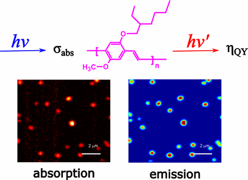
Figure 1. Photothermal microscopy in super critical liquid
- Wide-field photothermal microscopy: Current photothermal microscopy uses a confocal configuration, which severely limits the field of view and the time resolution when multiple nanoparticles must be addressed simultaneously, notably in biological systems. We aim to overcome the above limitations by wide-field photothermal microscopy. With collaboration of biology groups we aim to show the applicability of the new microscopy to live-cell imaging.
- Photothermal circular dichroism: Circular dichroism (CD) is the absorption difference between left and right circularly polarized light. The CD signal of molecules is typically very small and requires high sensitivity and careful control of the polarization to detect the tiny CD signal. We plan to apply photothermal detection to the CD signal of single metal nanoparticles, and ultimately of smaller and smaller amounts of chiral molecules.
- Mid-infrared photothermal microscopy: The absorption of mid-IR light by a molecule provides information about the molecule’s vibrational levels and can be used as a fingerprint of a molecule. We aim to use tunable mid-IR lasers as heating sources and a visible laser as the probe for photothermal imaging with chemical sensitivity.
References:
- Single pentacene molecules detected by fluorescence excitation in a p-terphenyl crystal, M Orrit and J. Bernard, Rev. Lett. 65 (1990) 2716-9.
- Hundreds-fold sensitivity enhancement of photothermal microscopy in near-critical xenon, X. Ding, L. Hou, H. V. Meer, A. P. Alivisatos, and M. Orrit, J Phys. Chem. Lett. 7 (2016) 2524-9.
- Absorption and quantum yield of single conjugated polymer poly[2-methoxy-5-(2-ethylhexyloxy)-1,4-phenylenevinylene] (MEH-PPV) molecules, L. Hou, S. Adhikari, Y. Tian, I. Scheblykin, M Orrit, Nano Lett. 17 (2017) 1575-81.